 English
 Français
 Deutsch
 Русский
 Italiano
 Español
 Português

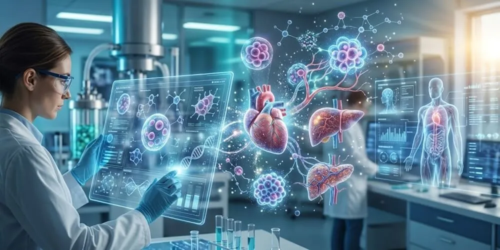
Stem cell-derived organ models are organized 3D cell cultures that recapitulate human organ physiology and disease pathology. They are generated from human pluripotent stem cells-embryonic stem cells or induced pluripotent stem cells-differentiated into specific cell types relevant to the organ of interest and assembled with biological or synthetic matrices providing support and spatial organization cues. Various combinations of the major human cell types found within each specific organ, together with inductive signaling and structural information provided by the environment, lead to the formation of functional organoid-like structures having at least some key organ-specific functions.
What is the concept of stem cell-derived organ models?
Stem cell-derived organ models have been able to predict accurately complex phenomena taking place within human organs which have not been replicable in conventional 2D cell cultures or animal experiments, such as toxicity induced by drugs, metabolism, and interaction between drugs. At present, most predictive value for drug effects in rodent models comes from detailed comparative characterization of human pathology; some information comes from studies using rodent organ models or other experimental rodents created for specific purposes. However, since stem cell-derived organ models do not replicate all aspects of any given organ their use is restricted to phenomena that involve exclusively the type of organ represented - such as rules governing transport, absorption, and excretion of drugs; receptor-ligand properties; viral tissue tropism; differences in metabolic pathways for specific drugs.
In what ways do stem cell-derived organ models differ from traditional models?
Key elements where cellular composition, physiological relevance, and data outcomes differ between stem cell-derived organ models versus traditional ones relate to these aspects. Traditional animal models are supposed to respond and translate into human disease, physiology, and pharmacology. Members of the biomedical community have long acknowledged that human biology cannot be exploited through animal species alone since they differ in anatomy, physiology, and regimens; species differences constrain or preclude certain tests as well as interpretation of testing and conclusions; trials on animals may serve poorly for safety indicators plus efficacy toward humans. In order to further knowledge about humans, models integrating complex biology into three-dimensional dynamic structures are urgently needed. Discovery pipeline facilitating systems should predict responses not found in animals or generate better estimates than comparisons between animal species; ideally, such systems would self-organize into tissue resembling organs displaying not only key structural features but also emerging behavioral responses associated with function.
The acceleration of the development process by 2027 will include three-dimensional dynamic human-relevant organ models that can provide information rapidly and accurately in the search for new therapeutic targets against major diseases such as cancer, neurological diseases, cardiovascular disease, and rare diseases. Primary organ-derived cells embedded in biocompatible matrices maintained under biomimetic conditions capable of supporting long-term physiological functions represent an important first step toward engineered organ-level models.
These approximations are crude and do not yet contain all the multiple cell types, vascularization, innervation, and interfacing with the immune system that real organs and tissues have. Therefore, there is still interest in developing organ-, organ-level-, and organ system-like structures with increasing complexity and physiological relevance. Progress toward complex 3D stem cell models is becoming possible through a better understanding of organogenesis and with the development of new culture surfaces and media that support multiple cell types and cell–cell interactions.
What are organoids in regenerative medicine? How are they used?
Stem cell-derived organ models differ from classical in vivo and in vitro alternatives because they better recapitulate human biology than animal models, share a genetic background with patients, and recreate dynamic aspects of organ function. Data throughput and ethical considerations are not as favorable compared to 2D models. Disease organoids that mimic the patient’s disease allow testing of patient-specific drugs and dosing, modeling toxicology and pharmacokinetics, as well as assessing immune responses resident in the organ.
Organoids are 3D stem cell-derived models used for regenerative medicine toward applications including cell therapy, disease modeling with patient-derived stem cells, and studies on pathophysiology. These models are different from systems since they do not contain all tissue types; therefore, supporting structures required for full vascularization, maturation, and integration into a host are missing from organoids. The typical workflow uses pluripotent stem cells as starting material, with controls for batch quality applied together with lineage-tracing strategies throughout differentiation.
How will 3D stem cell models shape research by 2027?
By 2027, it will be possible to use 3D stem cell models to advance research in every field substantially. New methods for fabrication will allow structural designs and compositional diversity never seen before! The performance of such models can now be quantified using existing imaging modalities along with omics technologies plus adaptive functional assays! Spatially aware vascularization, innervation, and immune components will further enrich these constructs!
Models of stem cells organized in 3D will hasten study in every area. The different methods to put things together—from natural grouping without scaffolds and using magne- or electrospinning to scaffolds that join or go away into tissue—will make it easier for more people to use. Special, custom, and ready-made systems for bioreactors will help culture go beyond simple still conditions. More tools in microscopy, transcriptomics, proteomics, metabolomics, lipidomics, glycomics, multiplexing of cytokines, DNA, RNA, and profiling chromosome shapes will give increasingly detailed spatially aware characterizations. As models get more sensitive, flexible, and demanding vascularization, innervation, and immune components will matter more. Models so enriched will label clones fluorescently, then test functional responses to perturbation by genetic or pharmacological means.
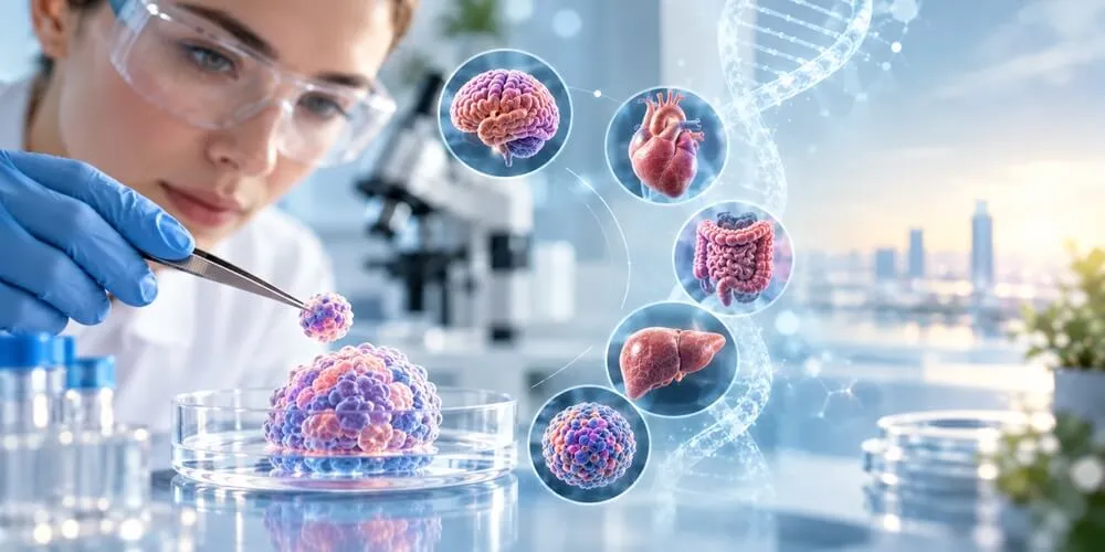
What advances drive disease modeling with stem cells today?
Three advances in stem cell biology are transforming disease modeling: the boom in human-induced pluripotent stem cell (iPSC) lines, fast and efficient genome editing with CRISPR/Cas9, and high-content screening of disease-associated iPSCs using multi-omics readouts. The availability of human iPSC panels opened avenues for high-throughput and patient-relevant drug discovery. Paradoxically, these advances address the major limitations of the traditional organotypic disease models.
Human-derived disease-specific cell types hold the promise of better cellular models, and the fruitful translation of basic stem cell-emerging concepts into disease modeling and therapeutic avenues demonstrates the capacity of the field to rapidly respond to current biomedical challenges. Human-derived iPSC lines of different genetic backgrounds combined with disease-agnostic CRISPR/Cas9 technology enable the generation of cellular models for genetic diseases on an unprecedented scale.
What ethical and regulatory factors guide organ and stem cell tissue models?
Ethical issues around these ways include how much like organs these organoids are when compared to adult organs and tissue; do they have enough complexity that they could achieve consciousness? Regulatory guidance is also a major aspect of this area of research. At present, medical products that involve organoids or similar 3D tissue structures represent new categories needing premarket approval. For products supporting human organ function, the FDA recommends for such developers establish a relationship between structure and function engineered tissues.
The use of stem cell-derived organ or tissue constructs for therapeutic purposes is expected to continue expanding, including applications in quality control, drug screening, disease modeling, testing new therapies, and predictive drug safety. Stem cell-derived mini-organs and tumor organoids that closely mimic real tumors will probably be used as the basis for predicting risks and responses in patients as well as developing preventive drugs and therapies. These models have even greater relevance in defining the physical and chemical microenvironments of the organs, autologous organ transplantation in regenerative and transplantation medicine, and drug testing platforms. By 2027, research into the practical application of tissue stem cells will likely merge with 3D printing technologies to create specific organ-like structures as a platform for long-term drug testing.
What will be the function of stem cell organoids 2027 in therapy development?
Organs and tissues derived from stem cells allow disease modeling, safety testing, and evaluation of biological plausibility. The outcomes can guide decisions on clinical use but require further steps for preclinical-to-clinical translation, including assessing physiological relevance to humans. This pathway is at the heart of parallel preclinical modeling of candidate responses before, during, and after clinical trials.
Organs and tissues derived from stem cells are increasingly developed in parallel with clinical trials. The first studies using these models to support preclinical development of therapies for patients with unmet clinical needs were reported in 2021; they verified patient-specific drug responses of solid tumors in organoid models prior to therapy and continued safety analysis on treatment afterwards. Results from an organoid study applied in parallel to a case of COVID-19 infection were published recently, involving preclinical modeling potential toxicities to the heart, kidney, and pancreas. Heterotopic implantation into liver by the patient’s neuroendocrine cancer expressing neuroendocrine features supported treatment decisions that was considered pioneering safety validation following direct administration.
Stem Cells Enable Creation of 3D Stem Cell Derived Tissue Models in Regenerative Medicine
Stem cell-derived tissue models have greatly advanced disease characterization. Multiple teams are working on standard operating procedures to make them. More specifically, the stem cell-derived vascular plexus for COVID-19 is an excellent model for studying infection by SARS-CoV-2, virus-induced pathogenesis, and testing therapeutic agents against such infections. Both stem cell tissue models can be used for planning experiments with transplantation and describing the pathogenesis of SARS-CoV-2. The heart vascular plexus derived from stem cells is also being utilized as a bridge to create functional myocardial tissue and its use in testing various cell sources and culture conditions for building myocardial constructs. A blood-brain barrier maintained on Transwell inserts is currently being used as a brain tissue model to study how peripheral inflammation affects the innate immune response to HIV-1 in brain macrophages. Moreover, protocols are under development for other brain organotypic cultures such as those of the hippocampus and fetal brain.
Protocols have been reported for several years in regenerative medicine regarding 3D cell type production of other organs. A recent comparison of three-dimensional (3D) retinal tissue with different cell origins on mechanism study of retinal pigment epithelium layer formation in RCS rats and an examination of X-linked retinoschisis mouse retina with multiple 3D retinal models support this direction. Three-dimensional normal artery models are reported to study the barrier function and drug-mediated modulation of blood-brain-blood-retina (BBBR) permeability and evaluate the safety and efficacy of treatment for retinitis pigmentosa. To address increasing perplexity concerning porous scaffolds in retinal fabrication, there is proposed utilization of mesoporous silica nanoparticles with dichroic light-up fluorescence.
What challenges appear in creating accurate models for complex organs?
Building frame models for complex organs is a challenging task, particularly when it comes to the brain, heart, and other large structures. The primary considerations are the maturity and functionality of the tissue. Organoids may mimic fetal and very early developmental stages of the organ, but such patterns need to be recapitulated later in life. Vascular networks supplying nutrients and oxygen as well as removing wastes are also difficult to establish due to changes with maturation and play crucial roles in some diseases too. Related to vascularization, the appropriate presence and function of immune cells during the reconstruction of the organ are critical for studying a wide range of human conditions. Engineering will assist with all these aspects using advanced tissue engineering facilities, 3D bioprinting, and microfluidic technologies but for now, the combination of stem cells with technology and materials cannot meet the demand for complex organs. A collaborative effort with the complementary field of bioengineering seems to be an appropriate pathway.
The maturation of organoids as well as their functional activity sometimes goes beyond what can be achieved within the field of stem cells. However, new developments in organ-on-chip micro- and multi-organ systems provide recent pathways for integrating lab models of organoids containing specific vascular tissues or organ systems (e.g., kidney splenic) to test drugs or corticosterone concerning human depressive disorders. Furthermore, social connection between vascular-tipped organoids has been reported to study normal neuron activity and its involvement in neuronal disease development and maturation.
How can institutions prepare for the future of stem cell research 2027?
Investments in core facilities that support 3D stem cell models will accelerate progress and empower labs. Basic training will equip researchers with essential skills while interdisciplinary teams combine expertise in stem cell biology, 3D printing or bioprinting, microfabrication, and tissue engineering. Institutional data infrastructures will meet the needs of new stem cell models, enabling storage and sharing access. Institutions will also review ethical committees to streamline, harmonize, and standardize pathways for stem cell research.
Pilot projects that align developmental programs with clinical research will facilitate integration into patient populations and clinical endpoints. Institutions should encourage collaborations where investigators from multiple labs agree to develop specific models for the same disease and share any resulting data; initiatives like these lay a foundation for data-sharing consortia that have volume, variety, and value needed to attract major funding.
Open data and standard formats will enable short-term advances in stem cell-derived organ modeling, while consortia will expedite long-term problems. Many groups are required to create and optimize models through intense collaboration, especially when they venture into new areas of research. Open data, released in standard formats, allow model developers to use outside datasets. Interoperability with data repositories aids resource conservation and permits independent discovery of new uses for existing resources or validation of prior results.
As stem cell models become more useful in applied settings, integration with industrial and regulatory partners will facilitate their routine use before transition to trial-ready platforms. Interdisciplinary efforts provide a combined skill and knowledge base for developing models of greater complexity as well as exploring diseases. The combination of cell types and physiological context—and ultimately whole-organ constructs—will increasingly reflect human biology under industry-standard doping and mutation profiles.
Stem cell–derived organ and tissue models are set to thrive in the coming years. Partnerships with external organizations and funding will help validate the models, while strategies that promote sharing and open access will enhance the generation of information as well as its use. These activities will close knowledge gaps, hence enabling successful translation into a broad range of therapeutic and screening applications.
How Organoids and Gene Editing Are Shaping the Future of Medicine
The effect of stem cell-derived organ and tissue models on biology as well as medicine is very great, likely to accelerate. Organs plus tissues made from stem cells have major advantages in biological relevance, predictive power plus scalability. Tissue fidelity will be increased soon; high-throughput applications will be enabled; systematic biological platforms for drug testing plus disease modeling will be created. Patient-derived iPSCs, CRISPR-based editing, multi-cellular tissue assemblies plus an open-data paradigm are key drivers here. Thus, it is realistic now to use these models for exploratory plus drug-discovery purposes—and developing regulatory pathways for their clinical application is a pressing aspiration.
The path toward clinical application is equally evident, with the promise of disease-modeling organoids being a major motivation. Organoids derived from patient tissues provide insights into mechanism and function, guide precision therapy design, and progress toward clinical application. Patient consent for organoids is easier than that needed for stem-cell-derived models. Coupled with genome-editing technologies plus the ability to recreate cellular complexity in vitro, these advances help dissect disease pathways plus associated drug-discovery opportunities.